A 5-month-old castrated male Smooth Fox Terrier presented for a 1-month history of right thoracic limb lameness.
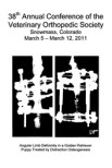
Physical examination revealed right elbow pain on extension and mild soft tissue swelling of the distal antebrachium. Radiographs and computed tomography showed elongated focal radiolucent regions in the distal radial metaphysis. There was incongruity of the right elbow with a short radius. Bone biopsy and histopathology of the regions confirmed a retained cartilaginous core characterised by bony trabeculae with frequently retained central cartilaginous cores.
A dynamic proximal ulnar ostectomy was performed to improve elbow congruity. The owner was instructed to restrict activity to short leash walks for 8 weeks followed by a gradual activity increase.
On follow-up examination 16 weeks after operatively, the lameness and elbow pain were resolved. Radiographs at that time showed a healed ulnar ostectomy, proper elbow congruity, and resolved retained cartilaginous core.